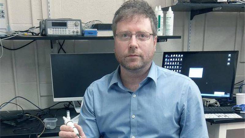
Dr. Jeremy Brown, QEII-affiliated scientist and associate professor with Dalhousie University’s School of Biomedical Engineering, recently received a five-year, $2.7-million grant to continue his groundbreaking work with microscopic endoscopes. This advanced medical technology uses ultrasound imaging and precision surgery to help doctors diagnose and treat life-threatening conditions such as blood clots and brain tumours.
Dr. Brown received his initial funding from ACOA in 2012. Within three years, the technology had been developed to the point of filing for patents, building prototypes and securing a licensing agreement. But for Dr. Brown, the main priority over the past three years has been to make the ultrasound “more sophisticated with a higher resolution and better frame rate” in order to improve its performance as a diagnostic tool and increase its potential value during live surgeries.
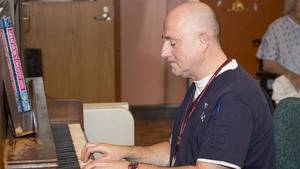
Designing the endoscope in the QEII's Centre for Clinical Research has provided Dr. Brown and his team with several unique advantages.
“The location is very convenient,” he says. “We have a lab in the Ear, Nose and Throat clinic and a lab in the medical sciences building across the street.”
When quizzed about the endoscope’s many applications, Dr. Brown becomes notably animated.
“The initial funding was for an endoscopic probe for the auditory system,” he says. “A partner company is using the ultrasound technology to manufacture and sell a probe to target imaging diagnostics inside the heart. And recently we picked up a new partner to use the same endoscope for neurosurgery.”
It’s this latest venture that has Dr. Brown particularly enthused.
“The neurosurgery component is going to be more focused on therapy,” he states. “We don’t just want to image tumours and guide the surgery; we also want it to cut the tumour.”
The high-resolution images produced by the device have already proven invaluable for the ENT clinic, but Dr. Brown maintains that the device’s true potential will be showcased in the operating room.
“Neurosurgeons are really pushing for it,” he says.
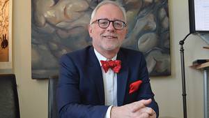
One of those neurosurgeons is Dr. Adrienne Weeks, who’s been watching this project with growing interest.
“It’s fabulous for neurosurgery,” she states. “We operate with less than millimetres between abnormal tissue and normal tissue. We can’t always see that, so, from a purely diagnostic standpoint, a high-resolution micro-ultrasound would be key.”
Dr. Weeks believes that this new method has “the potential to revolutionize our equipment and the way that we use surgical tools.”
“Current ultrasound probes are too large,” she notes. “They don’t fit in our small corridors and don’t give high enough resolution. You have the patient’s MRI but when you open the brain and start decompressing that tumour, your registration is immediately off because things move.”
A high-resolution miniature ultrasound would mitigate this, giving neurosurgeons the ability to gauge their status in real time.
“I can see exactly how far away I am from the brain stem,” Dr. Weeks illustrates. “And I know how much tumour is left.”
This equates to numerous advantages for QEII patients.
“Achieving a greater degree of resection translates to better outcomes and increased survival,” Dr. Weeks observes. “They experience less disability and return home sooner.”
An even more exciting prospect for Dr. Weeks is a treatment method called histotripsy, which future iterations of the endoscope will explore.
“Not only can I see something with high precision, I can use the ultrasound to vaporize that tissue through ablation without having to use suction or a scalpel,” she says.
Although additional research still needs to be done to compile diagnostic databases for various pathologies and determine how histotripsy will affect surrounding blood vessels in a live situation, Dr. Brown has every reason to be optimistic.
“It’s amazing how far they’ve come in just a few months,” says Dr. Brown.
This enthusiasm is mirrored by Dr. Weeks.
“I think this device has a potentially very large therapeutic benefit for patients, particularly in brain tumours,” she says with a hint of excitement. “Many of my colleagues are already saying ‘When is the ultrasound going to be ready? I want it!’”