One in eight men will be diagnosed with prostate cancer in their life, says Dr. Sharon Clarke, QEII radiologist in the Department of Diagnostic Radiology and assistant professor at Dalhousie University.
A digital rectal exam based on clinical suspicion has been the traditional method of detection. If the physician notices something out of the ordinary, a PSA, or Prostate Specific Antigen test, will usually follow.
“If that’s elevated, then they’ll go ahead and do a biopsy,” says Dr. Clarke.
This typically involves using an ultrasound transducer to visualize the patient’s prostate and remove 12 core samples, which are then sent for analysis. As Dr. Clarke explains, this approach can be problematic.
“It’s invasive and in some cases, may be painful or result in an infection.”
Dr. Clarke also notes that biopsies may detect slow growing, non-aggressive cancers that would never have caused any harm during the patient’s lifetime. This problem is called over diagnosis. A consequence of over diagnosis is that some patients may receive unnecessary treatments which may trigger a host of side effects. Even more problematic, Dr. Clarke states that biopsies “may outright fail to detect a clinically significant cancer.”
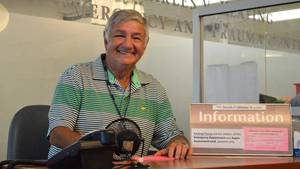
Michael Conde-Jahnel is well aware of these limitations. When Michael’s PSA reached a concerning level, his QEII urologist, Dr. David Bell, arranged for a series of biopsies. All of them came back negative.
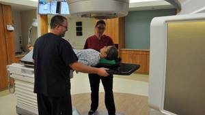
Nine months later, Michael’s PSA score increased, prompting Dr. Bell to send Michael for an extensive biopsy. When no health concerns were detected, Dr. Bell referred his patient to the team at BIOTIC, a core research facility at the QEII that works alongside clinical care, to use an innovative new non-invasive diagnostic test involving the new 3T Magnetic Resonance Imaging System. According to Dr. Clarke, MRI has become increasingly helpful in detecting and diagnosing prostate cancer.
“Our MRI research program has detected cancers in patients who had suspected cancer and numerous negative biopsies,” she says.
This was the case with Michael.
“I went through the MRI test and they confirmed that indeed there was some cancerous tissue there,” he says.
According to Dr. Clarke, the MRI method can show a more accurate reflection of a patient’s status.
“One particular patient’s biopsy showed low volume, low-grade cancer, but when we did the MRI, it showed that the cancer was more extensive and that it had spread outside the prostate gland. In addition, we can see things that help the surgeon determine or change their surgical approach to make sure that they get all the cancer.”
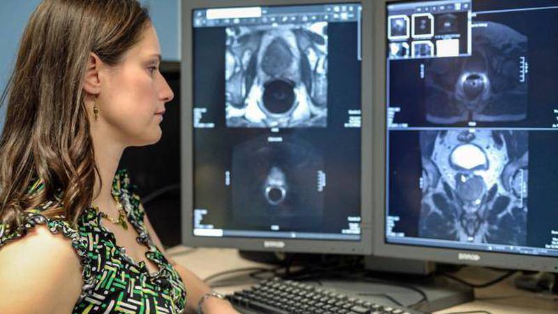
Critical to the MRI’s effectiveness are radiologists who scan through hundreds of images looking for anomalies. Given this daunting task, BIOTIC is now applying advanced imaging technology to help radiologists work faster and more intuitively.
“If you input all of these images into a mathematical model it will output two or three ‘maps’. The maps have condensed this information down and we feed this into our computer to help the radiologist figure out where the cancer is.”
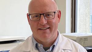
Research scientist Dr. James Rioux is applying this valuable data to help develop an automated computer-assisted classification algorithm to recognize prostate cancer.
“The idea is we’ll have a bunch of information from a number of different imaging contrasts,” he explains. “The computer knows where the cancer is based on radiologist findings. So it learns what features characterize cancer versus not cancer, benign versus malignant and so forth.”
Eventually the computer can be taught to extrapolate, or make an estimate from known facts.
“It can read in a new data set and calculate where the cancer is most likely to be,” he continues. “So the machine learning algorithm can go through and flag the most likely problem areas and tell the radiologist to have a closer look.”
Not only did this method prove to be definitive for Michael, he even describes the test as gentle and relaxing. After detection and treatment, he’s doing well.
“With me it’s a black-and-white situation. The cancer wasn’t discovered through conventional biopsy, it was the MRI diagnosis,” he maintains.
Dr. Clarke is quick to note this valuable asset wouldn’t be available without a major contribution from the QEII Foundation. $3.1-million in funding for the 3T MRI was provided by QEII Foundation donors, including a $2.5-million gift from the Gauthier and David families.
“It’s had a huge impact,” she says. “Because we have this 3T MRI, we have a research agreement with GE Healthcare and funding through the Atlantic Canada Opportunities Agency.”
With these resources now established, Dr. Clarke is hopeful for the future.
“We’re not developing these tools to be used just for us,” she indicates. “If we are successful then they can be further developed and marketed with GE’s software.”
The resulting technology has the potential to prolong and improve the lives of cancer patients around the world.
For Michael, this meant an early cancer diagnosis, allowing him to integrate the treatment as part of his daily life, including travel, consulting work and playing tennis several times a week – all at age 77.